What is a Baker’s Cyst?
In adults, Baker’s cysts behind the knee are usually caused by damage to the knee joint. They can cause mild pain and a feeling of tightness. Cysts often heal without treatment.
At a Glance
- A Baker’s cyst is caused by fluid accumulation in the knee cavity.
- It can cause mild pain and a feeling of tightness.
- In adults, cysts usually develop as a result of injuries or chronic joint diseases such as osteoarthritis or rheumatoid arthritis.
- Smaller cysts generally regress without treatment.
- Cooling and stretching the knee can relieve discomfort.
- If there are severe complaints, it is important to find and treat the cause of the cyst.
- Note: The information in this article cannot replace a doctor’s examination and should not be used for self-diagnosis or treatment.
What is a Baker’s Cyst?
A Baker’s cyst causes fluid to accumulate behind the knee. This leads to a protrusion in the knee joint capsule area.
The thigh and calf bones meet at the knee joint. There is a narrow joint space between them. This space is filled with fluid to keep the joint flexible. The joint and the joint space are surrounded by a joint capsule, which also contains a bursa.
A Baker’s cyst is an excessive accumulation of synovial fluid. This puts pressure on the posterior bursa, presenting as swelling behind the knee.
In adults, Baker’s cysts usually develop as a result of knee joint injury or inflammation. Depending on its size, the cyst often manifests as a feeling of tightness or pain in the knee.
Small Baker’s cysts often go unnoticed and heal without treatment. If symptoms occur, treatment is necessary.
Baker’s cysts are common in people over 50 or those with knee problems: approximately 5 to 40 out of 100 people with chronic knee pain have a Baker’s cyst. Baker’s cysts are rare in children.

What are the Symptoms of a Baker’s Cyst?
Larger cysts can cause the following discomfort:
- Tightness and a feeling of tension in the back of the knee
- Knee pain
- Stiff knee joint
- Swelling behind the knee, sometimes visible as a lump
- Swollen calf
- Swelling and pain that usually increases with movement of the knee
What Causes a Baker’s Cyst?
Baker’s cysts usually occur in adults after knee injuries, such as a meniscus tear, or as a result of chronic joint diseases like rheumatoid arthritis or osteoarthritis.
When the knee is damaged, it can no longer adequately absorb friction and impact. To compensate, more synovial fluid is produced in the joint capsule. This thick, clear body fluid nourishes the cartilage in the knee joints and acts as a “joint lubricant.” Excess synovial fluid is pushed into the bursa behind the knee, causing it to expand and form a Baker’s cyst.
If the cause is a condition like acute inflammation in knee osteoarthritis, and the condition resolves on its own, the Baker’s cyst may also regress without treatment.
What Factors Contribute to the Formation of Baker’s Cysts?
Over the years, the stability of the knee joint capsule decreases. Overuse or strain can cause cracks in the capsule. This leads to fluid exchange between the joint space and the bursa, triggering the development of Baker’s cysts.
Older adults are more likely to have had previous knee injuries. Chronic joint diseases also become more common with age. Both contribute to the formation of Baker’s cysts.
What Complications Can Arise from Baker’s Cysts?
If the cause of the Baker’s cyst is treated and, as a result, less joint fluid is produced, the cyst will regress. However, if the underlying condition is not effectively treated, Baker’s cysts can persist for years.
A Baker’s cyst can sometimes burst (rupture). In this case, joint fluid leaks out and spreads into surrounding tissues such as the calf muscles. This usually results in sudden, severe pain in the knees and calves. Bruising (hematoma) may also occur. The leaking fluid will gradually break down again. Sometimes, the tissue becomes inflamed after the Baker’s cyst bursts. Therefore, it is advisable to see a family doctor.
If the cyst presses on blood vessels, fluid accumulation (edema) may occur, causing swelling in the calf. If the Baker’s cyst presses on nerves, it can cause numbness and muscle weakness in the calf. However, such complications are rare.
How is a Baker’s Cyst Diagnosed?
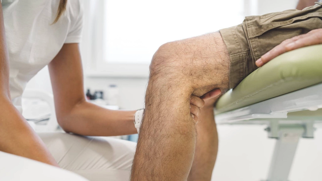
A Baker’s cyst is best observed when the affected leg is stretched. The doctor can then see and feel the fluid retention as a solid lump.
When the knee is bent, the cyst relaxes and softens or disappears completely.
If a definitive diagnosis cannot be made, further investigations are needed. Doctors then use imaging techniques such as:
- Ultrasound
- Magnetic resonance imaging (MRI)
These methods make it possible to detect changes in the joints or tissues. For example, small cysts can be detected during an ultrasound examination.
How is a Baker’s Cyst Treated?
Baker’s cysts are only treated if there are symptoms. For example, elevating the leg after exertion, using bandages, or applying pain-relieving ointments can be tried as self-help measures.
Anti-inflammatory painkillers and physical therapy can also be helpful. The goal of physiotherapy is to keep the knee flexible and stable. This involves specific leg muscle exercises.
An orthopedic specialist can aspirate the cyst or the knee joint. This involves inserting a needle into the cyst or knee joint and withdrawing the fluid. Surgery is also an option if severe symptoms cannot be relieved by other means, but only if it can treat the cause of the Baker’s cyst.


